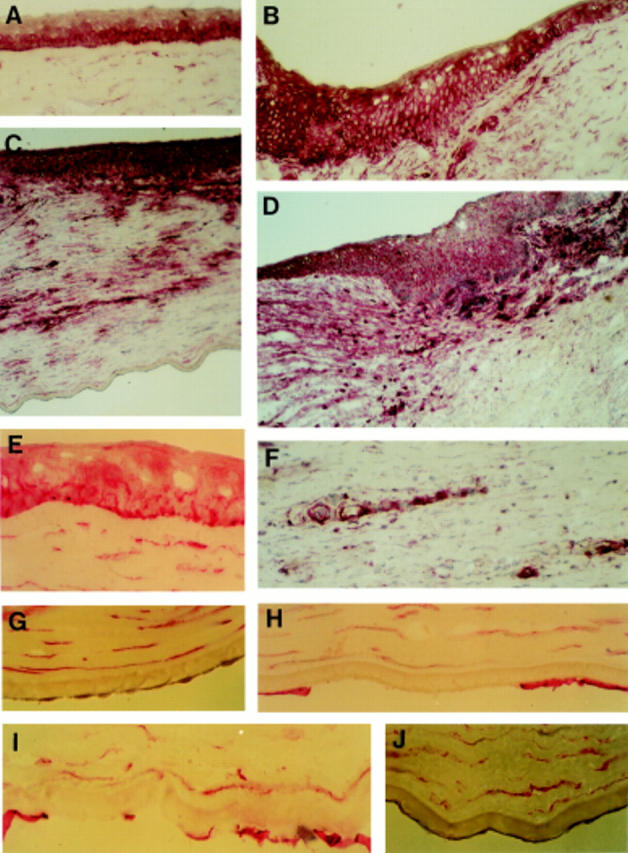
Figure 1 .

Alkaline phosphatase anti-alkaline phosphatase immunostaining for CD44 in human corneal sections. Original magnification × 200 for (A), (B), (C), (D) and (F); and × 400 for the others. (A) Epithelial cell staining in normal central region; (B) epithelial cell staining in normal limbus; (C) section from a cornea with keratitis showing intense staining in all epithelial layers, but no staining on endothelial cells; (D) section from a cornea with allograft rejection showing the staining of epithelium in the region where graft and receptor corneas adjoin; (E) epithelial cell staining in Fuchs' endothelial dystrophy; (F) CD44 staining on neovascular endothelium and infiltrating cells in cornea with keratitis; (G) normal corneal endothelial cells showing no staining for CD44; (H) section from a cornea with pseudophakic bullous keratopathy showing positive staining on the remaining endothelial cells; (I) section from a cornea with Fuchs' endothelial dystrophy showing the CD44 staining on the remaining endothelial cells; and (J) section from a cornea with keratoconus showing negative staining of endothelial cells for CD44.
