
Figure 2 .
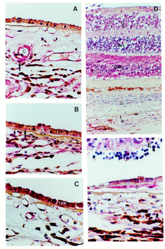
Light microscopic images of immunohistochemistry with anti-TGF-β (A and B), b-FGF (C), and PDGF-AA (D and E) antibodies. (A) Control maculae show TGF-β expression in smooth muscle cells (long arrow) and endothelial cells (short arrow) of choroidal blood vessels (C). The RPE shows only faint staining in most of the control maculae. (B) TGF-β expression is increased in the RPE of this typical ARM macula. Basal laminar deposit (long arrow) contains TGF-β as well. Note that the most prominent RPE staining is present on top of a drusen (open arrow) that itself shows no staining. (C) A typical example of an ARM macula that shows b-FGF expression in the RPE. (D) PDGF-AA expression in a macula with neovascular macular degeneration. The retina demonstrates PDGF-AA expression, whereas the retina in control maculae shows no staining at all. Note that the expression in the outer nuclear layer (ONL) is more prominent than in the inner nuclear layer (INL) and ganglion cell layer (G). (E) PDGF expression in a large drusen (D) in a macula with neovascular macular degeneration. S = scar tissue, b = Bruch's membrane. Original magnification × 400.