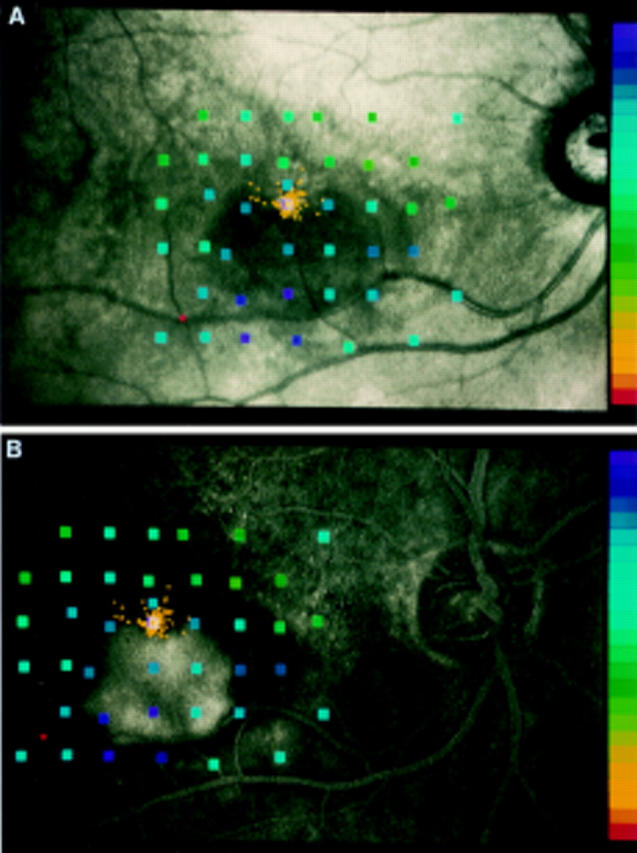
Figure 2 .

Results of static fundus perimetry in a right eye of a patient with choroidal neovascularisation (CNV). (A) Standard image with threshold values according to the right hand scale with brightest stimuli at the top and smallest contrast between stimuli and background at the bottom. Single fixation points are drawn as yellow dots. (B) Results converted onto the mid time sequence of fluorescence angiography which enables direct visualisation of mean fixation point in relation to classic CNV.
