
Figure 4 .
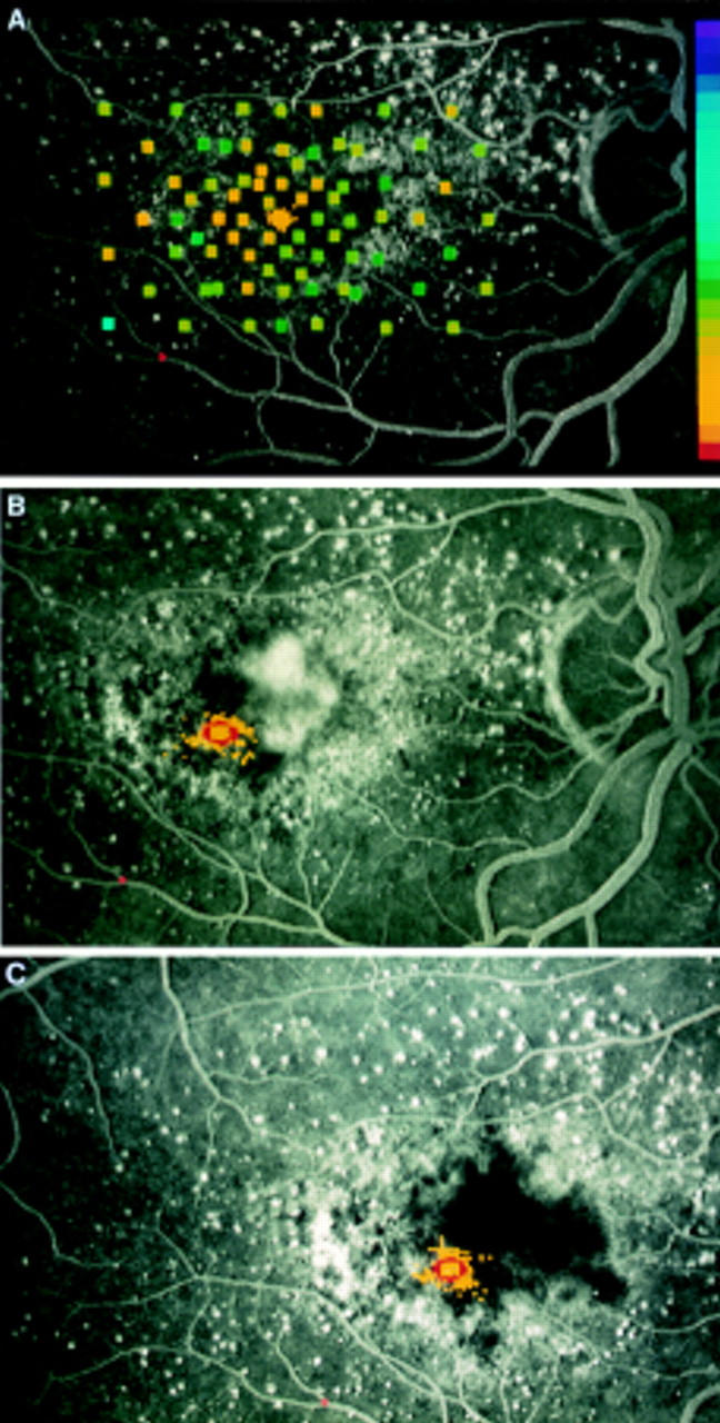
Right eye of a 67-year-old woman first presenting with decrease of visual acuity in the left eye. Right eye showed nearly normal perimetric results with the SLO (right hand scale with brightest stimuli at the top and smallest contrast between stimuli and background at the bottom) and central fixation (yellow dots) while angiography demonstrates macular degeneration without defined leakage (A). (B) Six months later juxtafoveolar CNV nasally of the fovea can be seen during fluorescein angiography, mean fixation point has shifted temporally and stability of fixation is worse (red circle). (C) Four months after laser treatment no recurrence can be seen, visual acuity is 0.4, and reading ability is improved subjectively. Location and stability of fixation as measured during SLO perimetry are about the same.