Abstract
BACKGROUND—Most idiopathic macular holes can be closed by a surgical procedure combining vitrectomy, posterior hyaloid ablation, and fluid-gas exchange followed by postoperative positioning. Reopening of closed macular holes has been reported, but its frequency is not known. Here the incidence of reopening after successful macular hole surgery is reported. METHODS—77 consecutive cases of idiopathic macular holes operated with autologous platelet injection between July 1993 and October 1995 were reviewed. The procedure consisted of three port vitrectomy, posterior hyaloid removal, non-expansile fluid-gas exchange, and autologous platelet injection followed by face down positioning. The incidence of reopening was analysed in the cohort of the 72 anatomical successes. RESULTS—Mean follow up was 12.3 months. The macular hole reopened in five eyes of five patients (five out of 72 patients, 6.9%), in four cases after cataract extraction. In four cases too, an epiretinal membrane was noted, either clinically or during reoperation, and fluorescein leakage in the macular area was present in two cases. Three of the five cases of reopening were reoperated and all three were anatomical successes. CONCLUSION—Late macular hole reopening occurred in five out of 72 patient, and in four cases after cataract surgery. The presence of an epiretinal membrane around the hole in four of them suggested that tractional forces were responsible for the reopening. Reoperation, performed in three cases, again closed the macular holes.
Full Text
The Full Text of this article is available as a PDF (189.2 KB).
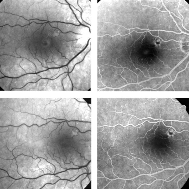
Figure 1 .
Case 1, a full thickness stage 3 macular hole before and after operation. Top left, red-free photograph. At presentation, macular hole with elevation of edges. Visual acuity (VA) was 20/60. Top right, fluorescein angiography. Faint central hyperfluorescence of the hole (note also the pre-existent extramacular subretinal pigmentation). Bottom left, red-free photograph. Closure of the macular hole 1 month after surgery. VA=20/30. Bottom right, fluorescein angiography. The central hyperfluorescence has disappeared.
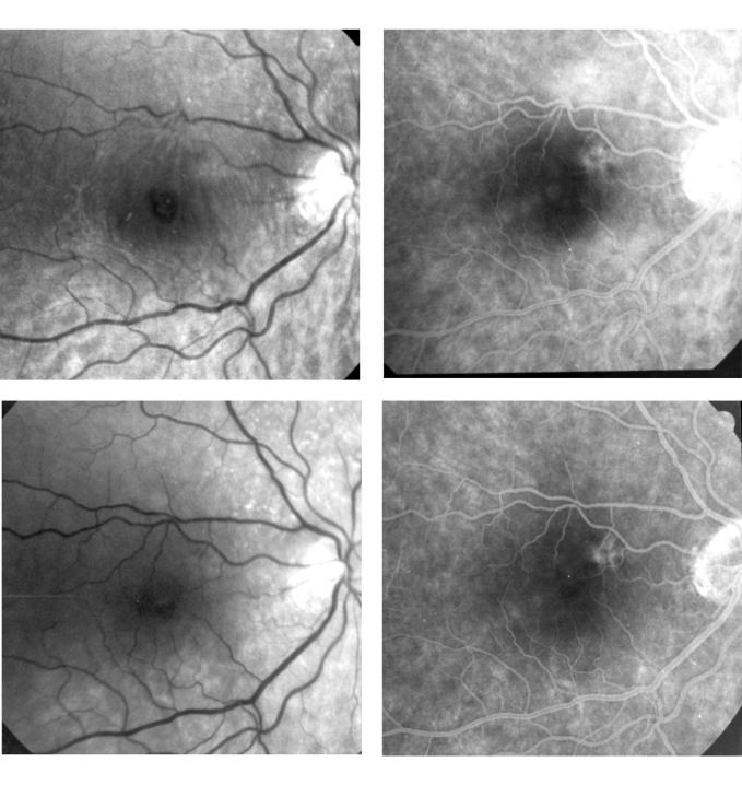
Figure 2 .
Case 1. Macular hole reopening after cataract extraction, and closure after reoperation. Top left, blue filter photograph: reopening of the macular hole with epiretinal membrane formation 7 months after cataract surgery and 19 months after macular hole surgery. VA=20/125. Top right, fluorescein angiography. Round central hyperfluorescence of the hole and moderate intraretinal dye leakage, with no cystoid spaces in the macula. Bottom left, red-free photograph. Closure of the hole after reoperation and complete removal of the epiretinal membrane. VA= 20/50. Bottom right, fluorescein angiography showing faint residual hyperfluorescence around the macula.
Selected References
These references are in PubMed. This may not be the complete list of references from this article.
- Duker J. S., Wendel R., Patel A. C., Puliafito C. A. Late re-opening of macular holes after initially successful treatment with vitreous surgery. Ophthalmology. 1994 Aug;101(8):1373–1378. doi: 10.1016/s0161-6420(13)31174-9. [DOI] [PubMed] [Google Scholar]
- Ezra E., Arden G. B., Riordan-Eva P., Aylward G. W., Gregor Z. J. Visual field loss following vitrectomy for stage 2 and 3 macular holes. Br J Ophthalmol. 1996 Jun;80(6):519–525. doi: 10.1136/bjo.80.6.519. [DOI] [PMC free article] [PubMed] [Google Scholar]
- Fekrat S., Wendel R. T., de la Cruz Z., Green W. R. Clinicopathologic correlation of an epiretinal membrane associated with a recurrent macular hole. Retina. 1995;15(1):53–57. doi: 10.1097/00006982-199515010-00010. [DOI] [PubMed] [Google Scholar]
- Funata M., Wendel R. T., de la Cruz Z., Green W. R. Clinicopathologic study of bilateral macular holes treated with pars plana vitrectomy and gas tamponade. Retina. 1992;12(4):289–298. doi: 10.1097/00006982-199212040-00001. [DOI] [PubMed] [Google Scholar]
- Gass J. D. Idiopathic senile macular hole. Its early stages and pathogenesis. Arch Ophthalmol. 1988 May;106(5):629–639. doi: 10.1001/archopht.1988.01060130683026. [DOI] [PubMed] [Google Scholar]
- Gass J. D. Reappraisal of biomicroscopic classification of stages of development of a macular hole. Am J Ophthalmol. 1995 Jun;119(6):752–759. doi: 10.1016/s0002-9394(14)72781-3. [DOI] [PubMed] [Google Scholar]
- Gaudric A., Massin P., Paques M., Santiago P. Y., Guez J. E., Le Gargasson J. F., Mundler O., Drouet L. Autologous platelet concentrate for the treatment of full-thickness macular holes. Graefes Arch Clin Exp Ophthalmol. 1995 Sep;233(9):549–554. doi: 10.1007/BF00404704. [DOI] [PubMed] [Google Scholar]
- Gaudric A., Massin P., Qinyuan C. An aspirating forceps to remove the posterior hyaloid in the surgery of full-thickness macular holes. Retina. 1996;16(3):261–263. doi: 10.1097/00006982-199616030-00016. [DOI] [PubMed] [Google Scholar]
- Glaser B. M., Michels R. G., Kuppermann B. D., Sjaarda R. N., Pena R. A. Transforming growth factor-beta 2 for the treatment of full-thickness macular holes. A prospective randomized study. Ophthalmology. 1992 Jul;99(7):1162–1173. doi: 10.1016/s0161-6420(92)31837-8. [DOI] [PubMed] [Google Scholar]
- Gordon L. W., Glaser B. M., Ie D., Thompson J. T., Sjaarda R. N. Full-thickness macular hole formation in eyes with a pre-existing complete posterior vitreous detachment. Ophthalmology. 1995 Nov;102(11):1702–1705. doi: 10.1016/s0161-6420(95)30806-8. [DOI] [PubMed] [Google Scholar]
- Kelly N. E., Wendel R. T. Vitreous surgery for idiopathic macular holes. Results of a pilot study. Arch Ophthalmol. 1991 May;109(5):654–659. doi: 10.1001/archopht.1991.01080050068031. [DOI] [PubMed] [Google Scholar]
- Le Gargasson J. F., Rigaudiere F., Guez J. E., Gaudric A., Grall Y. Contribution of scanning laser ophthalmoscopy to the functional investigation of subjects with macular holes. Doc Ophthalmol. 1994;86(3):227–238. doi: 10.1007/BF01203546. [DOI] [PubMed] [Google Scholar]
- Lewis H., Cowan G. M., Straatsma B. R. Apparent disappearance of a macular hole associated with development of an epiretinal membrane. Am J Ophthalmol. 1986 Aug 15;102(2):172–175. doi: 10.1016/0002-9394(86)90139-x. [DOI] [PubMed] [Google Scholar]
- Madreperla S. A., Geiger G. L., Funata M., de la Cruz Z., Green W. R. Clinicopathologic correlation of a macular hole treated by cortical vitreous peeling and gas tamponade. Ophthalmology. 1994 Apr;101(4):682–686. doi: 10.1016/s0161-6420(94)31278-4. [DOI] [PubMed] [Google Scholar]
- Melberg N. S., Thomas M. A. Visual field loss after pars plana vitrectomy with air/fluid exchange. Am J Ophthalmol. 1995 Sep;120(3):386–388. doi: 10.1016/s0002-9394(14)72169-5. [DOI] [PubMed] [Google Scholar]
- Oshika T., Yoshimura K., Miyata N. Postsurgical inflammation after phacoemulsification and extracapsular extraction with soft or conventional intraocular lens implantation. J Cataract Refract Surg. 1992 Jul;18(4):356–361. doi: 10.1016/s0886-3350(13)80071-5. [DOI] [PubMed] [Google Scholar]
- Park S. S., Marcus D. M., Duker J. S., Pesavento R. D., Topping T. M., Frederick A. R., Jr, D'Amico D. J. Posterior segment complications after vitrectomy for macular hole. Ophthalmology. 1995 May;102(5):775–781. doi: 10.1016/s0161-6420(95)30956-6. [DOI] [PubMed] [Google Scholar]
- Poliner L. S., Tornambe P. E. Retinal pigment epitheliopathy after macular hole surgery. Ophthalmology. 1992 Nov;99(11):1671–1677. doi: 10.1016/s0161-6420(92)31746-4. [DOI] [PubMed] [Google Scholar]
- Wendel R. T., Patel A. C., Kelly N. E., Salzano T. C., Wells J. W., Novack G. D. Vitreous surgery for macular holes. Ophthalmology. 1993 Nov;100(11):1671–1676. doi: 10.1016/s0161-6420(93)31419-3. [DOI] [PubMed] [Google Scholar]


