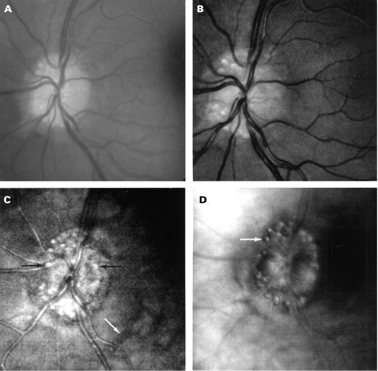
Figure 1 .

Fundus images of a patient with optic disc drusen. (A) Fundus photograph showing superficial disc drusen. (B) Short wavelength (540 nm, green) SLO images showing superficial structures including disc drusen, retinal vessels, and the nerve fibre layer. (C) Long wavelength (780 nm, infrared) confocal mode SLO images showing more drusen including buried ones (black arrows) which are not visible in (A) or (B). Note the retinal vessels appear out of focus as they lie superficial to the plane of focus, whereas deep choroidal vessels are now visible (white arrow). (D) Indirect mode (780 nm, infrared) SLO images of disc drusen showing the distinctive `bubble' appearance.
