
Figure 1 .
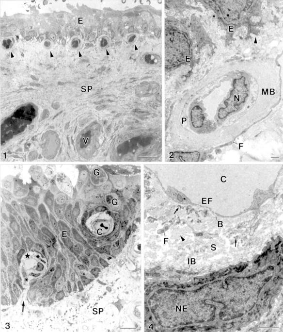
(1) The stroma of the pterygium (SP) is highly vascularised (V). Subepithelial capillary (arrowhead), epithelium (E). Bar = 20 µm. (2) Subepithelial capillary. Subepithelial basal lamina (arrowhead), epithelial cell (E), cellular extension of a fibroblast (F), multilamellar basal lamina of a capillary (MB), endothelial cell nucleus (N), pericyte (P). Bar = 1 µm. (3) Intraepithelial capillary (C) and a capillary with a hilus-like connection (arrow) between the perivascular connective tissue (star) and the subepithelial stroma of pterygium (SP). Fibroblast (asterisk), goblet cell (G), epithelium (E). Bar = 10 µm. (4) The perivascular stroma (S) of intraepithelial capillaries (C) is outlined by an endothelial basal lamina (B) and an "internal" epithelial basal lamina (IB). The perivascular stroma contains cellular processes of fibroblasts (F), collagen fibrils (arrowhead) and electron dense elastin-like material (arrow). Endothelial cell fenestration (EF), epithelial cell nucleus (NE). Bar = 1 µm.