
Figure 2 .
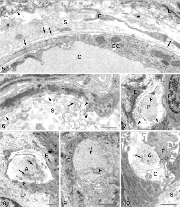
(5) The endothelial basal lamina of the intraepithelial capillaries (C) is partially single layered and partially multilayered (arrow). Anchoring fibrils (arrowhead), wide spaced collagen fibrils (asterisk), epithelial cell (E), endothelial cell (EC), extension of fibroblast (F), perivascular stroma (S). Bar = 1 µm. (6) Defects in the "internal" epithelial basal lamina (arrowhead). Anchoring fibril (arrow), epithelial cell (E), perivascular stroma (S). Bar = 0.5 µm. (7) Defects (arrowhead) in the epithelial basal lamina at a stromal micropapilla (P). Epithelial cell (E), extension of fibroblast (F). Bar = 1 µm. (8-10) The enlarged intercellular space within the epithelium contains structures not typical for epithelia with collagen fibrils (arrow) and fibroblast processes (F). Amorphous material (A), non-identified cell (C), epithelial cell (E), stroma (S). Bar = 1 µm.