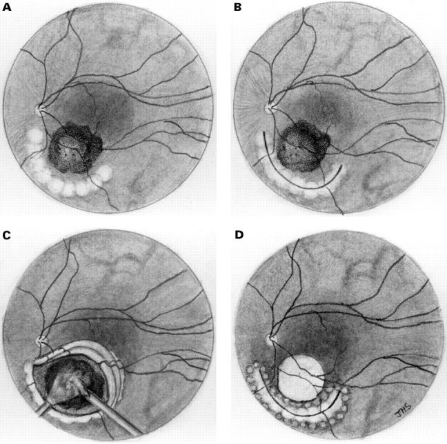
Figure 1 .

Schematic diagram illustrating the salient features of the surgical technique. (A) Endodiathermy is applied in an arcuate fashion to the retina peripheral to the tumour. (B) A retinotomy is created with intraocular scissors and dissected free of the tumour in an anterior to posterior direction. (C) Diathermy and argon laser endophotocoagulation are applied over the tumour surface and margins and, under hypotensive anaesthesia, all visible tumour is resected with the vitrector down to bare sclera. (D) The retinal flap is reposited and an air-fluid exchange performed. Laser is applied to the edges of the retinotomy and the eye is filled with silicone oil (5000 centistokes).
