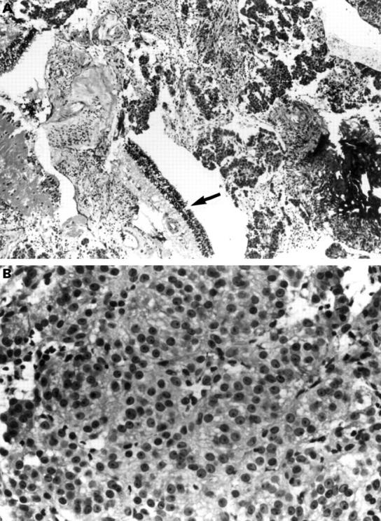
Figure 5 .

(A) Histological section of the material collected and spun down from the vitrectomy cassette demonstrating clumps of epithelioid melanoma cells together with bits of normal retina (arrow) and choroid (haematoxylin and eosin, original magnification, ×100). (B) Higher power view of a clump of tumour cells consisting almost exclusively of epithelioid melanoma cells (haematoxylin and eosin, original magnification ×400).
