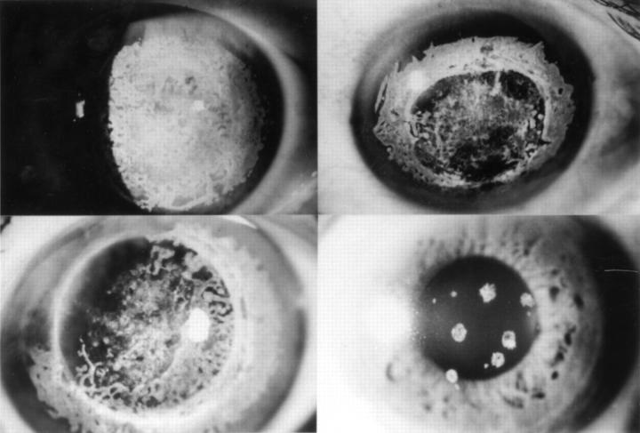
Figure 4 .
Slit lamp photograph of the corneas of recurrence. Untreated recipient corneas show dense subepithelial opacities in each case. Top left, case 3, 29 years after the second operation performed at the age of 21. Top right, case 4, 20 years after the first operation at the age of 25. Bottom left, case 5, 21 years after the second operation at the age of 53. Bottom right, daughter of case 5 at 48 years of age. Cornea shows only several granular opacities.

