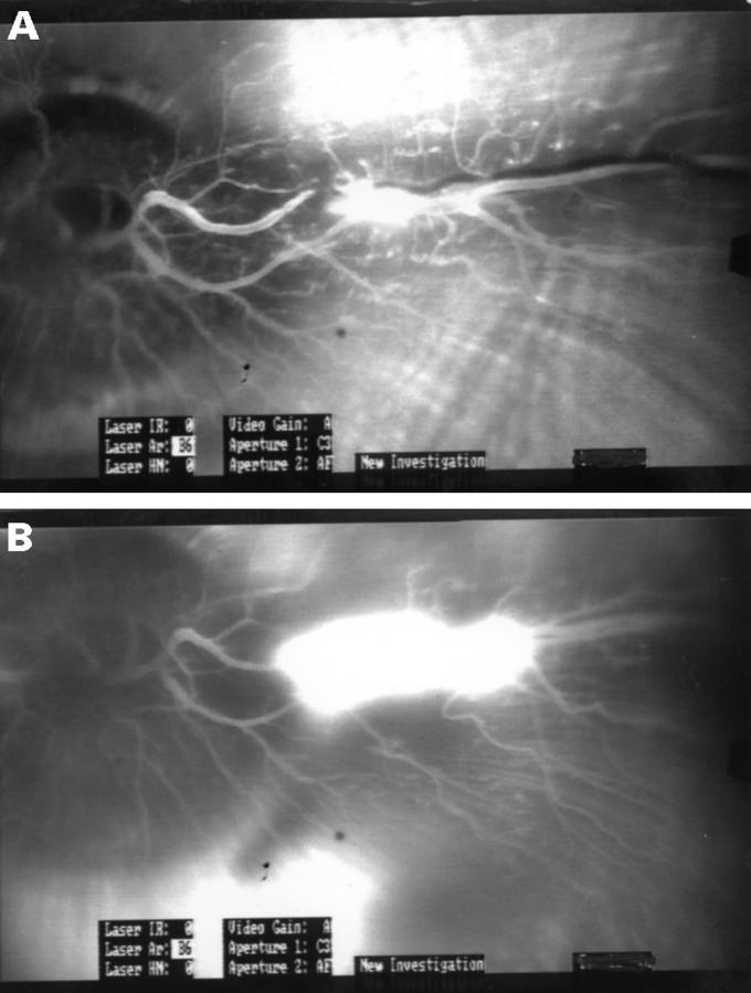
Figure 1 .
Theses fluorescein angiograms show an eye from group 3 that had been treated with both streptokinase and ultrasound. (A) The angiogram 30 minutes after the laser treatment shows that the vein is totally occluded peripheral to the thrombus. There is fluorescein in that part of the vein that is central to the thrombus, because of small veins that empty in the central part of the vein. (B) The angiogram 12 hours after the treatment shows that the vessel is open. Leakage from the retina of the fluorescein is noted around the place where the laser treatment had been performed.

