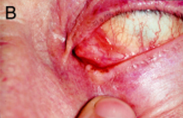
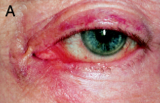
Figure 1 .
(A) Appearance of left lower eyelid before incisional biopsy. The contour is a distorted by the fibrotic lesion at the medial end of the lid. It is pale with telangiectatic vessels and has indistinct margins. The tumour has no cilia on it. (B) The tumour involves the full thickness of the lid including the posterior lamella. It surrounds the lower punctum and has obliterated its lumen.


