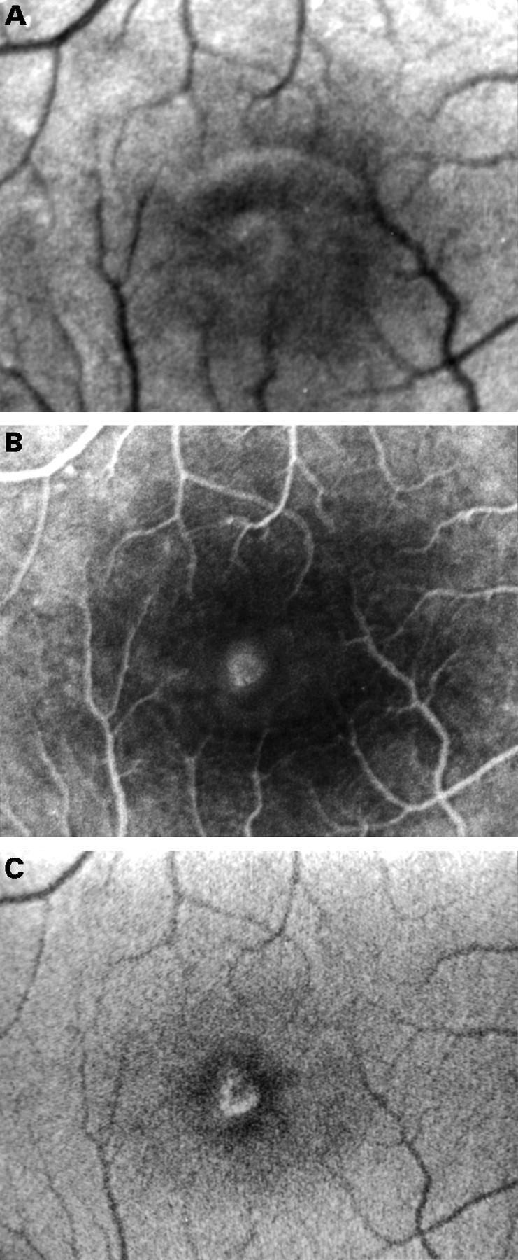
Figure 2 .

Fundus photograph of the right eye with a stage 2 full thickness macular hole (A). Fluorescein angiography shows a small window defect (B). Autofluorescence imaging shows increased fluorescence centrally. The less fluorescent zone within the macular hole in the superior-nasal part may represent a partially attached operculum (20° field) (C).
