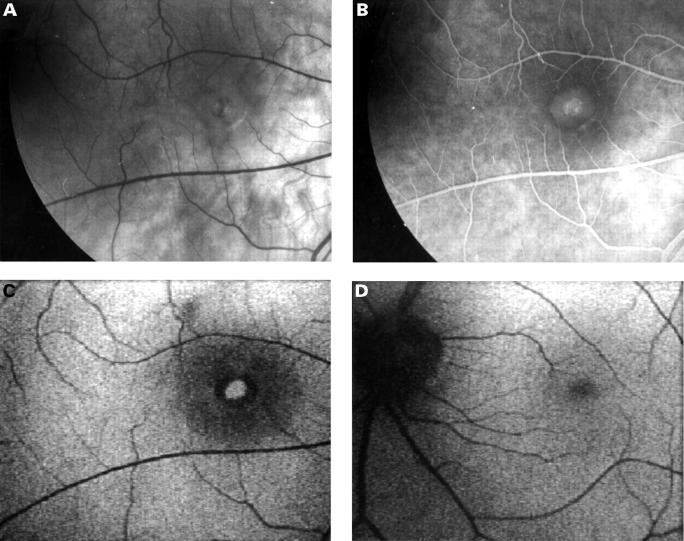
Figure 3 .
Fundus photograph and fluorescein angiogram of the right eye with a stage 4 full thickness macular hole (A, B). Autofluorescence imaging shows increased autofluorescence centrally corresponding to the macular hole and a ring of decreased autofluorescence corresponding to the cuff of subretinal fluid. The shallow subretinal fluid which extends beyond the cuff is seen as an area of decreased autofluorescence surrounding the cuff of the hole (40° field) (C). The contralateral normal eye shows even autofluorescence (D).

