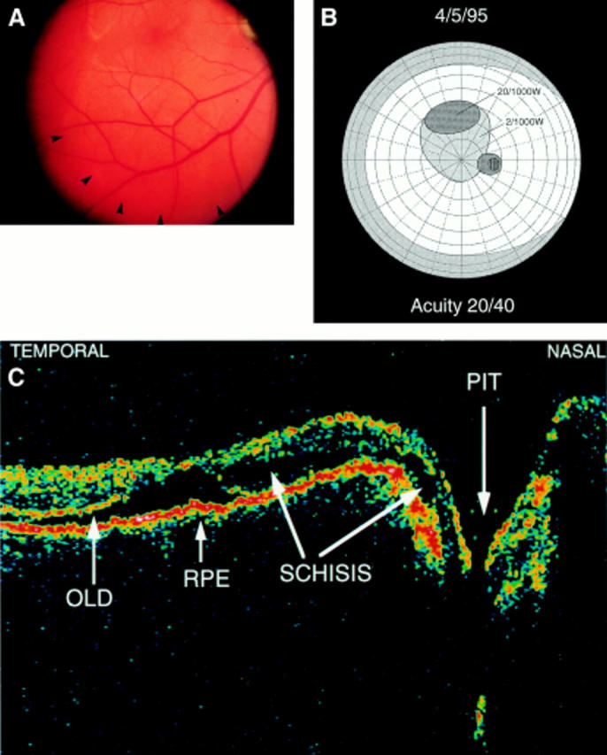
Figure 3 .

(A) Posterior pole (patient 1) at 3 weeks. The OLD is displaced below the inferior temporal vein (arrowheads). (B) Central visual field at 3 weeks. It demonstrates a dense scotoma (20/1000W) between 15° and 25° above fixation consistent with the OLD. A less dense scotoma (2/1000W) persists in the posterior pole. (C) OCT at 3 weeks. It reveals the retinal layers almost attached to the pigment epithelium. A hole in the OLD at the macula is apparent. The schisis-like cavity persists and is confluent with the optic disc pit. RPE = retinal pigment epithelium; OLD = outer layer detachment; − horizontal section through macula and disc.
