
Figure 2 .
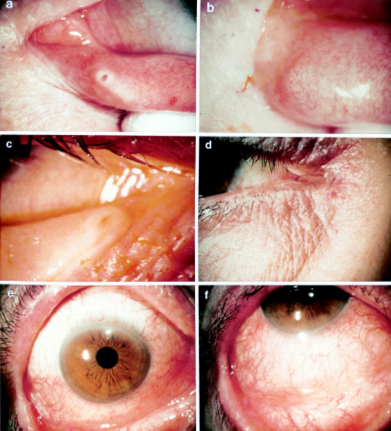
External photographs of a representative case with delayed tear clearance showing no apparent obstruction at the punctum (a). The corresponding fluorescein clearance test (FCT) is shown in Figure 1(g) and (h). For comparison, another representative case with delayed tear clearance showing punctal obstruction (b), for which the corresponding FCT is shown in Figure 1(i) and (j). The remaining photographs illustrate the typical appearance of a swollen punctum yielding a slit-like appearance (c), the subcutaneous oedema of the adjacent skin (c, d), bulbar conjunctival injection preferentially in the inferior aspect (e), and tarsal conjunctiva with a mild papillary response (f).