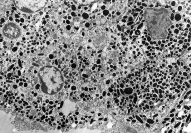
Figure 2 .
An electron micrograph of a melanocytic hamartoma in the uveal tract of TPras transgenic mice. Both spindle cells and cuboidal cells with oval nuclei containing marginated heterochromatin are identified. Intracytoplasmic membranous vesicles, premelanosomes, phagosomes, and cigar-shaped and oval melanin granules are identified. The melanosomes measure between 0.4 and 1.50 µm. Atypical nuclear features are identified in one cell (lower left) which contains a prominent nucleolus (original magnification ×5510).

