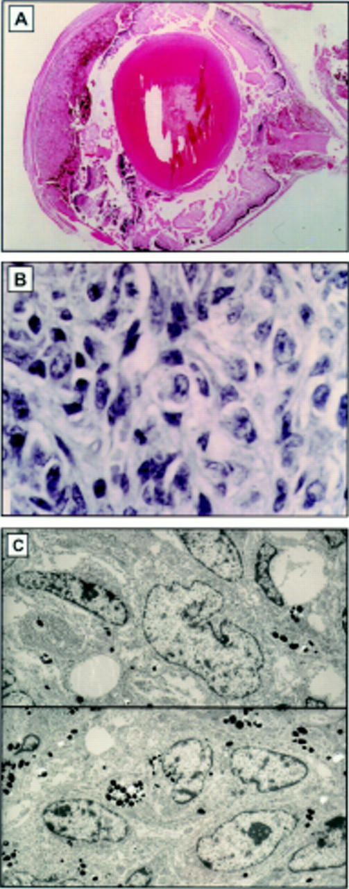
Figure 3 .

(A) Light micrograph of melanoma in the uveal tract of TPras transgenic mice. The melanocytic proliferation is seen to fill the entire uveal tract. Cells are moderately pigmented and morphological features are not readily discernible (haematoxylin and eosin, original magnification ×10). (B) High power light micrograph of the bleached specimen demonstrates a proliferation of pleomorphic, spindle-shaped cells. The cytological characteristics of the malignant melanoma in the 3 week TPras mouse are demonstrated. (C) Ultrastructurally, the cells are spindle shaped with oval to round nuclei, increased nuclear to cytoplasmic ratios, marginated chromatin, irregular nuclear envelope, and prominent nucleoli. Intracytoplasmic melanin granules are demonstrated (haematoxylin and eosin, original magnification ×160).
