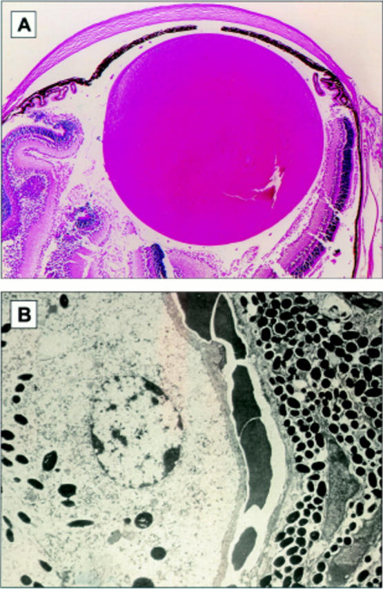
Figure 4 .

(A) Photomicrograph of the anterior uveal tract in one of the control (negative littermate) mice. The iris ciliary body and choroid are pigmented and normal in structure. There is no evidence of infiltration hyperplasia or hypertrophy of the uveal tract (original magnification ×44). (B) Electron micrograph demonstrating the retinal/choroidal junction in one of the control mice. Choroidal melanocytes are spindle-shaped cells with oval nuclei containing a bland chromatin pattern. Intracytoplasmic round and oval melanin granules are identified. The melanosomes measure 0.50 µm by 0.75 µm. The RPE is composed of cuboidal cells with oval nuclei containing marginated heterochromatin. There is apical to basal polarity and basement membrane formation. Intracytoplasmic rough endoplasmic phagosomes and membranous vesicles are identified. Intracytoplasmic cigar-shaped and oval melanin granules are identified and measure 0.44 µm by 1.58 µm (original magnification ×9180).
