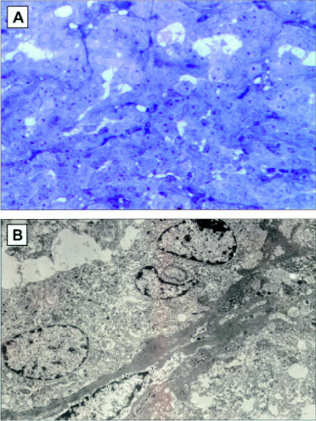
Figure 5 .

(A) Light micrograph demonstrating a retinal pigment epithelial adenomatous proliferation found in the uveal tract of a normal control litter mate. (B) Histopathologically, this was characterised as a melanotic proliferation of RPE between the retina and the choroid. The cells were noted to form tubuloacinar configurations (haematoxylin and eosin, original magnification ×160).
