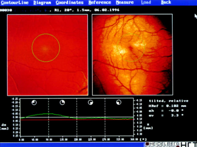
Figure 1 .
HRT scan of a normal macula in a healthy 36 year old man. The height variation of the contour line is shown in green and the reference plane (in red) is adjusted to the lowest point of the contour line. The scan is centred at the fovea. The circle centre is the fovea, the diameter is 2 mm. The scale (25 mm) is given on the left hand vertical axis.

