
Figure 4 .
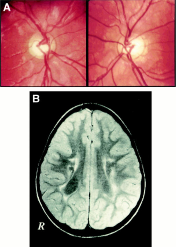
(A) Fundus photograph of patient no 9 demonstrating large cups in normal sized discs. (B) This single MRI shows typical changes of periventricular leucomalacia with atrophic dilatation of the posterior parts of the lateral ventricles. The amount of periventricular white matter is reduced around the occipital horns and adjacent to the trigone bilaterally. Remaining white matter has an abnormally bright signal on this T2 weighted image, first echo, indicating permanent damage, gliosis. Variations of signal involving left posterior cortical structures represent artefacts in the image.