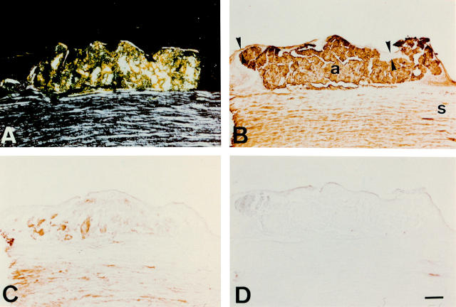
Figure 2 .
Immunostaining of gelatinous drop-like corneal dystrophy cornea with anti-apolipoprotein J and anti-apolipoprotein E antibodies. The epithelium (delineated by arrowheads in (B)) varies greatly in thickness. (A) When viewed under polarised light, congo red staining is seen in epithelial and subepithelial regions, showing a birefringent polarisation typical of amyloid. The posterior cornea is not visible here because tissue was obtained by a lamellar keratoplasty. (B) The antibodies to apolipoprotein J stain the subepithelial amyloid deposit (a) fairly intensely, and the stroma (s) more diffusely. (C) The anti-apolipoprotein E antibody also stains amyloid, but much less strongly than the anti-apolipoprotein J antibodies. (D) Control section incubated with normal mouse serum IgG shows no discernible immunolabelling. Bar=100 µm.

