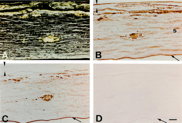
Figure 3 .
Immunostaining of a lattice corneal dystrophy type I cornea with anti-apolipoprotein J and anti-apolipoprotein E antibodies. The epithelium is delineated by arrowheads, and the endothelium identified by an arrow. (A) When viewed under polarised light, congo red staining is seen subepithelially and in corneal stroma, showing birefringent polarisation typical of amyloid. (B) Immunostaining for apolipoprotein J clearly mirrors the distribution of subepithelial and intrastromal amyloid deposits. (C) Apolipoprotein E immunoreactivity also co-exists with amyloid, but appears to stain the section less strongly than the apolipoprotein J antibodies. (D) Control section incubated with normal mouse serum IgG shows no discernible immunolabelling. Bar=100 µm.

