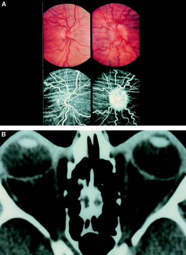
Figure 2 .

(A) Bilateral choroidal folds and unilateral papilloedema. Colour and fluorescein photographs demonstrating bilateral coarse choroidal folds and unilateral papilloedema in a patient (patient no 32, Table 3) with benign intracranial hypertension. (B) Distended nerve sheath and compressed globes. Computed tomograph scan of the same patient demonstrating dilated optic nerve sheaths. This patient presented with acquired hypermetropia. Examination revealed bilateral choroidal folds and a swollen left disc. The arrow denotes perineural cerebrospinal fluid.
