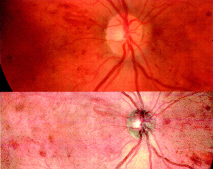
Figure 2 .

Upper image is a conventional fundus camera image of a patient with mild diabetic retinopathy. The lower image is the colour SLO picture of the same patient. Note that for the colour SLO image two separate images have been combined to give the requisite field of view.
