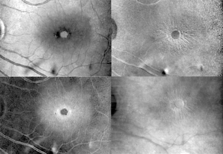
Figure 4 .
Patient with a macular hole. The top left is an SLO image of the fundus using an infrared laser; the top right shows a topographic image in which white indicates elevated areas; bottom left is a thickness image in which white indicates thicker areas; and bottom right is an indirect mode image.

