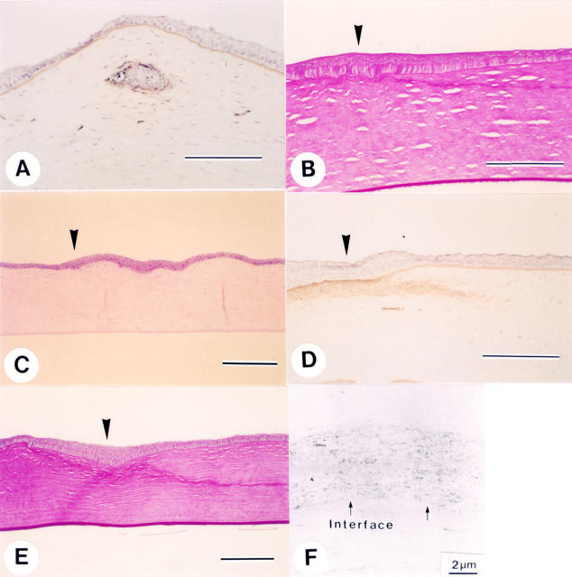
Figure 2 .
(A) Positive staining for type IV collagen is seen around the epithelial ingrowth portion on day 7 after LASIK (bar 200 µm). (B) PAS positive material is noted along the lamellar interface. Arrowhead indicates the wound edge (postoperative month 1, bar 200 µm). (C) Haematoxylin and eosin staining section 3 months after LASIK showing almost normal appearing cornea. Arrowhead indicates the wound edge (bar 200 µm). (D) Diffuse staining for type IV collagen is seen beneath the epithelium at the wound margin. Arrowhead indicates the wound edge (postoperative 3 months, bar 200 µm). (E) PAS stained section 9 months after LASIK. Note that the PAS positive material still present along the interface. Arrowhead indicates the wound edge (bar 200 µm). (F) Electron microscopy at 9 months after LASIK. Disorganised extracellular matrix is deposited along the interface (arrows).

