
Figure 2 .
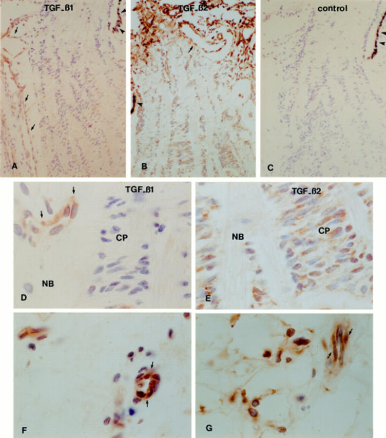
TGF-β1 and TGF-β2 immunoperoxidase staining in fetal optic nerve head (gestational age 24 weeks). TGF-β1 immunoreactivity was restricted to blood vessels throughout the optic nerve head (A, D) and TGF-β2 reactivity was localised to the extracellular matrix and cells in the optic nerve head (B, E). In the sclera, TGF-β1 was detected in association with blood vessels (F) and TGF-β2 was associated with the vessels and fibroblasts (G). (C) Illustrates the negative control. Arrows point to blood vessels and arrowheads point to the retinal pigment epithelium. NB = nerve bundles; CP = cribriform plates. (A-C) ×75; (D-G) ×750.