
Figure 4 .
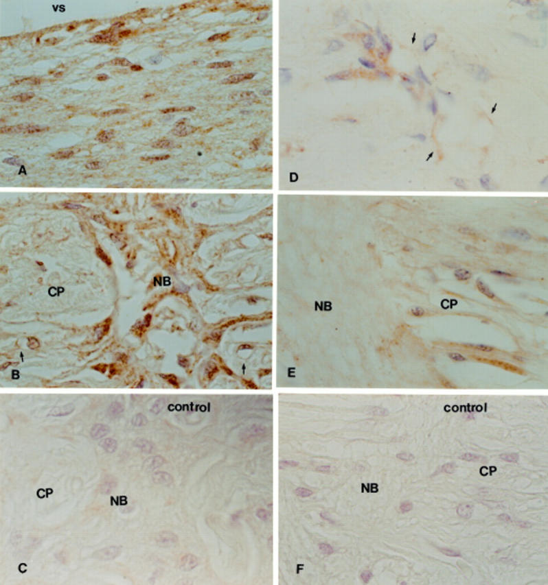
TGF-β2 immunoperoxidase staining in advanced (A, B, and C) and mild (D, E, and F) primary open angle glaucoma, donor ages 74 years old (advanced glaucoma) and 67 years old (mild glaucoma). Note widespread staining of astrocytes and the extracellular matrix throughout the optic nerve head of the sample from with advanced glaucoma (A and B). Reactivity in blood vessels was also noted (arrow). In the mild glaucoma sample, few astrocytes and blood vessels (arrows) were labelled in the prelaminar region (D) and in the lamina cribrosa, with little staining in the extracellular matrix (E). (C) and (F) are negative controls. vs = vitreal surface; NB = nerve bundles; CP = cribriform plates. ×750.