
Figure 1 .
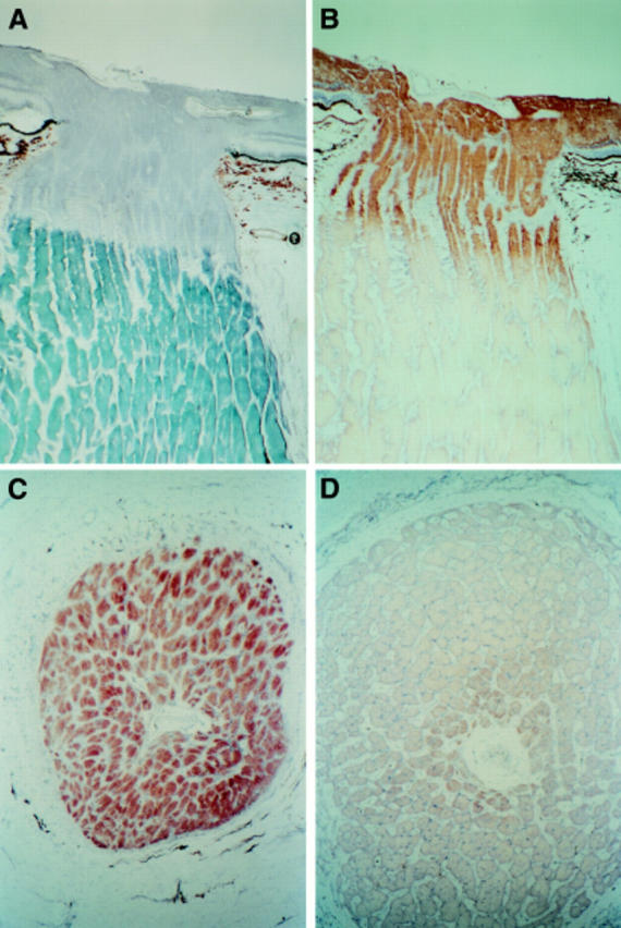
Photomicrographs of longitudinal sections through the optic nerve (A) stained with Sudan black to demonstate myelin and (B) reacted to demonstrate COX which is confined to the unmyelinated portion of retinal ganglion cell axons. Transverse sections of optic nerve (C) in immediate prelaminar zone showing high COX activity and (D) in the retrolaminar zone approximately 1.5 mm from section (C) showing low COX activity.