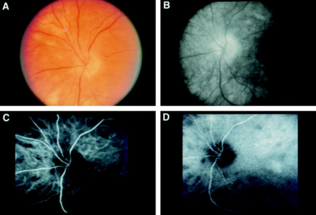
Figure 1 .
(A) Photograph of the left peripapillary fundus of a patient with short standing birdshot retinochoroidopathy, showing relatively few birdshot lesions and optic disc oedema. (B) Late phase of fluorescein angiography of the same eye, demonstrating silence of birdshot lesions. Optic disc leakage also is evident. (C) and (D) Early and late phases respectively of indocyanine green angiography of the same eye, revealing more birdshot lesions than ophthalmoscopy or fluorescein angiography. The lesions manifest early in the study and persist essentially unchanged. There is no sign of the optic disc oedema evident with fluorescein angiography.

