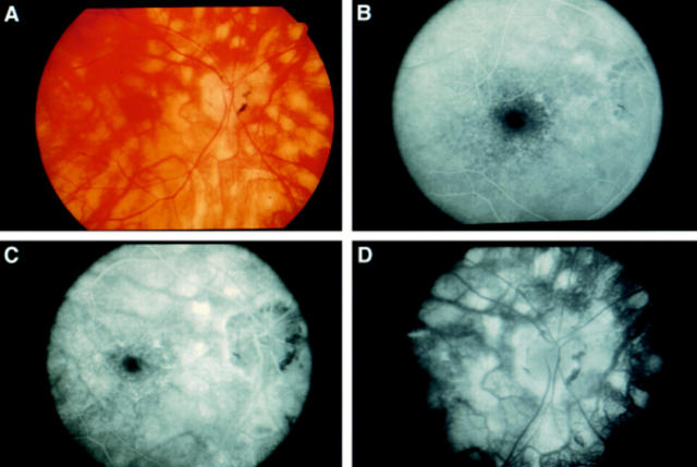
Figure 2 .
(A) Photograph of the right posterior pole of a patient with long standing birdshot retinochoroidopathy, showing diffuse birdshot lesions and circumpapillary retinal atrophy. (B), (C), and (D) Early, mid, and late phases respectively of fluorescein angiography of the same eye, demonstrating staining of birdshot lesions.

