
Figure 6 .
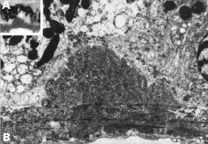
Preclinical drusen, entrapment site. This drusen appeared hyalinised on light microscopy (inset A) but EM (B) shows that it is an entrapment site composed of coated membrane bound bodies. A 50 year old man with normal fundal appearance, vision 6/6. (A) ×500, (B) ×5600.