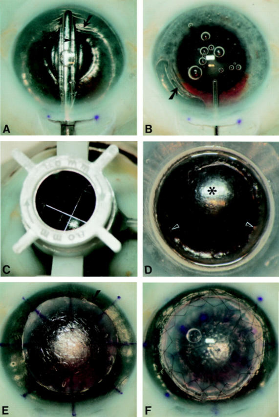
Figure 3 .

Demonstration of the surgical technique in a human eye bank eye. (A) The pocket is dissected first across the vertical meridian, and then extended sideways up to the limbus over 360°, with the same spatula. Note that the anterior chamber is completely filled with air, and that the dissection depth can be monitored by the width of the dark band (arrowhead) in between the spatula and the air to endothelium light reflex. Note also the wrinkling of the posterior corneal tissue (arrow). (B) After most air has been removed from the anterior chamber, the stromal pocket is filled with viscoelastic. Note the step ladder configuration of the relaxed posterior corneal tissue (arrow) which is pushed downward. (C) After trephination with a Hessburg-Barron trephine (D) an anterior corneal lamella is excised. Note the smooth recipient bed (asterisk) with some residual posterior stroma overlying the pupillary border (arrowheads). Pupillary dilatation was not intended as a part of the procedure. (E) After stripping Descemet's membrane, a "full thickness" donor button (arrow) is placed onto the recipient bed, and the donor and recipient corneal surfaces are marked with an eight incision radial keratotomy marker. (F) The donor button sutured in place with two running sutures. Pupillary dilatation was not an intended part of the procedure.
