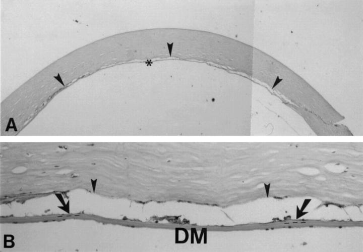
Figure 4 .
Light microscopy of a deep lamellar dissection through a scleral incision in a human eye bank eye. (A) A deep stromal dissection level (arrowheads) is seen (98% corneal depth). (B) Few stromal lamellae (arrows) are visible between the stromal dissection and Descemet's membrane; the dotted line indicates the junction of the stroma and Descemet's membrane (haematoxylin and eosin, original magnification ×35 and ×450).

