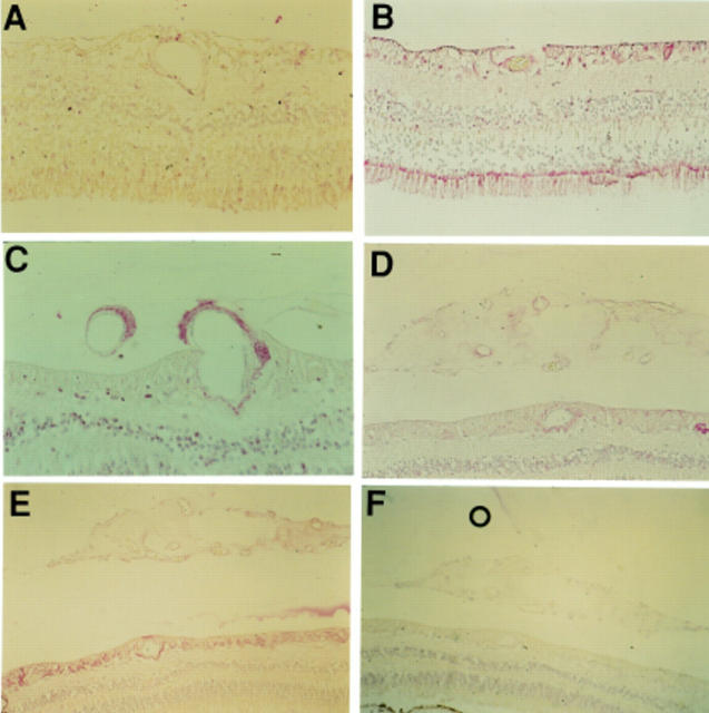
Figure 4 .
Photomicrographs demonstrating FLT-4 staining of normal retina (A), diabetic retina with obvious vascular intraretinal changes but no evidence of PDR (B), diabetic retina with PDR (C, D) and the same retina stained with GFAP (E). Immunostaining for FLT-4 was raised in diabetic tissue compared with non-diabetic tissue. Immunostaining was intermediate in the PDR specimens. Immunoreactivity for FLT-4 was abolished in a control specimen of PDR retina processed with omission of the primary antibody (F). Magnification A, D ×156; B ×118; C ×94; E, F ×60.

