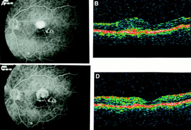
Figure 3 .
Multifocal choroidopathy. (A) Fluorescein angiography before surgery (VA= 20/40). (The broken line on FA frame indicates the OCT scan direction). (B) OCT before surgery: the neurosensory thickness is 475 µm; a focal hyperreflectivity lying above the band corresponding to the RPE can be visualised. (C) Fluorescein angiography after surgery (VA= 20/30): the CNV has been completely removed. (The broken line on FA frame indicates the OCT scan direction). (D) OCT after surgery: the normal foveal profile is restored and retinal thickness is 160 µm. The hyperreflectivity interpreted as the CNV is no longer visible.

