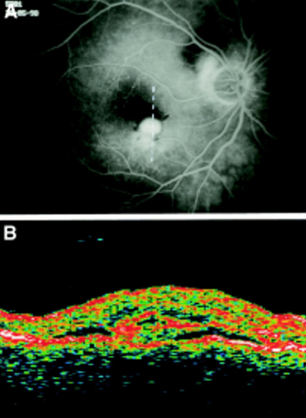
Figure 4 .

AMD complicated by CNV (OCT pattern A). (A) Fluorescein angiography shows a juxtafoveal and a peripapillary CNV. (The broken line on FA frame indicates the OCT scan direction). (B) The OCT scan performed through the juxtafoveal CNV reveals a marked thickening (503 µm) of the neurosensory retina and an increased reflectivity above the RPE interpreted as the CNV. A thin optically empty space, signifying neurosensory detachment, surrounds the CNV.
