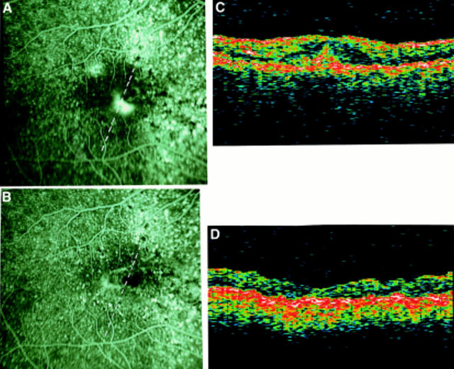
Figure 7 .
AMD complicated by subfoveal CNV. (A) Fluorescein angiography before surgery (VA=20/200). (The broken line on FA frame indicates the OCT scan direction). (B) The OCT clearly displays a focal hyperreflectivity lying above the RPE interpreted as the CNV. In this case the OCT shows the relation between CNV and the centre of the fovea. The adjacent neuroretina presents a non-homogeneous, reduced, hyperreflectivity due to oedema. The retinal thickness is 379 µm. (C) Fluorescein angiography after surgery (VA= 20/60). The loss of retinal pigment epithelial cells and choriocapillaris is minimal. (The broken line on FA frame indicates the OCT scan direction). (D) The focal hyperreflectivity identified by OCT before surgery is no longer visible. The retinal thickness is 154 µm.

