
Figure 2 .
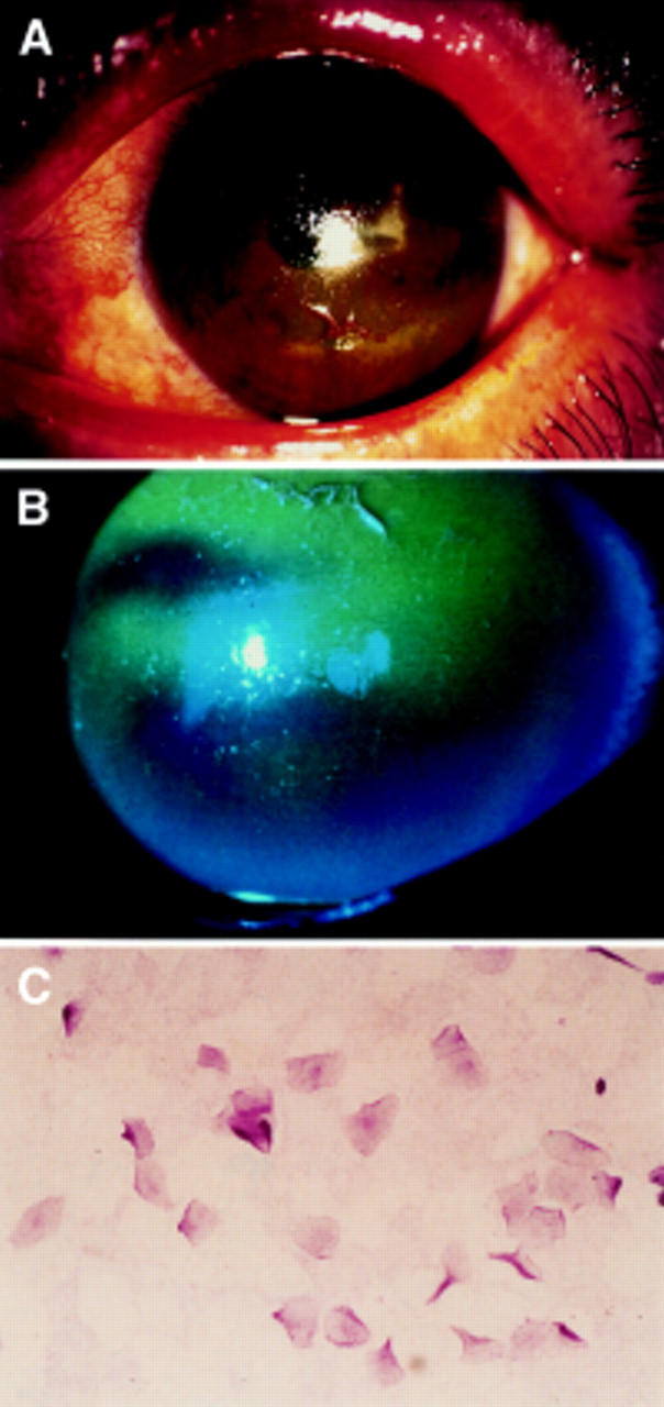
Slit lamp photographs stained by rose bengal and fluorescein of the left eye of the patients described (A, B). Note the corneal epithelium was deeply stained by fluorescein. Impression cytology of the bulbar conjunctiva in patients with Sjögren's syndrome (C) stained by PAS staining. There are no goblet cells observed. The epithelial cells are enlarged showing the squamous metaplasia.