
Figure 3 .
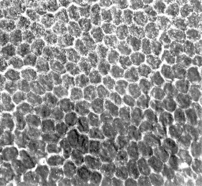
Scanning electron microscopy. Microvilli were found in abnormally high numbers on the surface of many endothelial cells (upper half of the photograph).
Official websites use .gov
A
.gov website belongs to an official
government organization in the United States.
Secure .gov websites use HTTPS
A lock (
) or https:// means you've safely
connected to the .gov website. Share sensitive
information only on official, secure websites.
Scanning electron microscopy. Microvilli were found in abnormally high numbers on the surface of many endothelial cells (upper half of the photograph).