
Figure 4 .
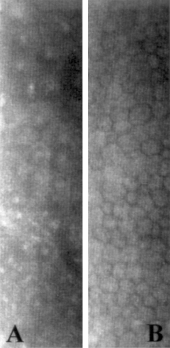
Specular microscopy. (A) Rounded dark cells with light borders and occasional light bodies within cell boundaries could be identified in the endothelium of the abnormal cornea. (B) In the fellow eye, the endothelium exhibited polymegathism, but no characteristics of ICE syndrome.