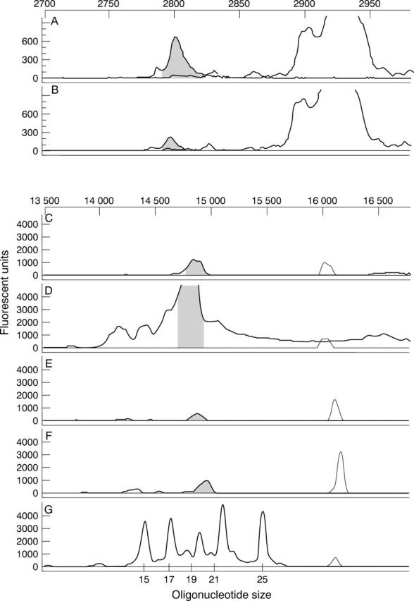
Figure 4 .

Persistence of PS-ODN following intravitreal injection demonstrated by Genescan analysis. Electrophoretograms of DNA extracted from the retina (A, C, and E) and the RPE-choroid-sclera complex (B, D, and F) at 6 weeks (A, B), 8 weeks (C, D), and 12 weeks (E, F) after injection with DS 012, and the oligonucleotide size standard (G). The internal standard is superimposed in all electrophoretograms at the same data collection point and the peaks for the PS-ODN are equivalent for both tissue samples at all time points. Note the actual fluorescent intensity of 6 week samples is effectively 130 times higher than that of 8 and 12 week samples.
