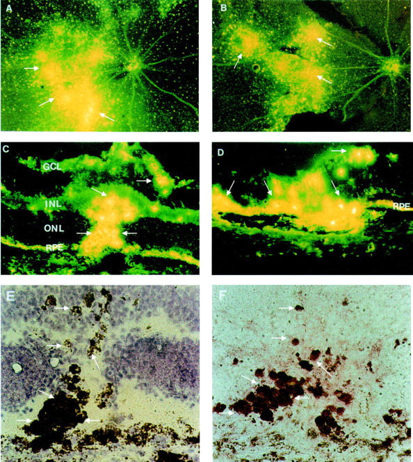
Figure 5 .

Distribution of PS-ODNs (DS 012, 5.0 nmol) in the retina following krypton laser photocoagulation. (A, B) Retinal whole mounts, 4 weeks (A) and 8 weeks (B) after injection; the PS-ODNs are concentrated in the regions of laser photocoagulation with an appearance of clumps of bright granules at the sites of laser burns (arrows). (C, D) Cryosections from eyes injected with DS 012 following krypton laser photocoagulation, 4 weeks (C) and 8 weeks (D) after injection. The fluorescent signal is particularly localised to infiltrating cells (arrows) and the RPE layer. (E) Higher magnification of (C) observed by light microscopy, counterstaining with haemotoxylin, the arrows indicating pigment laden infiltrating cells. (F) A serial section of (C) and (E), immunostaining for CD68 (arrows). Original magnification: (A, B) ×10; (C, D) ×50; (E, F) ×100. GCL = ganglion cell layer; INL = inner nuclear layer; ONL = outer nuclear layer; RPE = retinal pigment epithelium.
