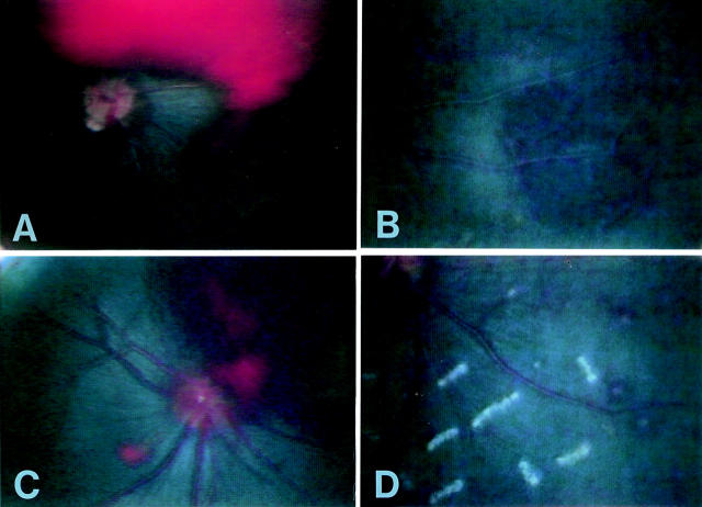
Figure 2 .
Haemorrhagic changes. (A) Vitreous haemorrhage—was seen in two of the 20 gerbils from day 7 after infection of T canis. This was associated with vitreous opacity and was absorbed within 1 month. (B) Choroidal haemorrhage—was found in 95% of gerbils with a single oral inoculation of T canis embryonated eggs. (C) Retinal haemorrhage—was seen in 55% of T canis infected gerbils. Large central retinal haemorrhage was observed. (D) White centred retinal haemorrhage. Multiple small haemorrhage with a white central part were found in the periphery.

