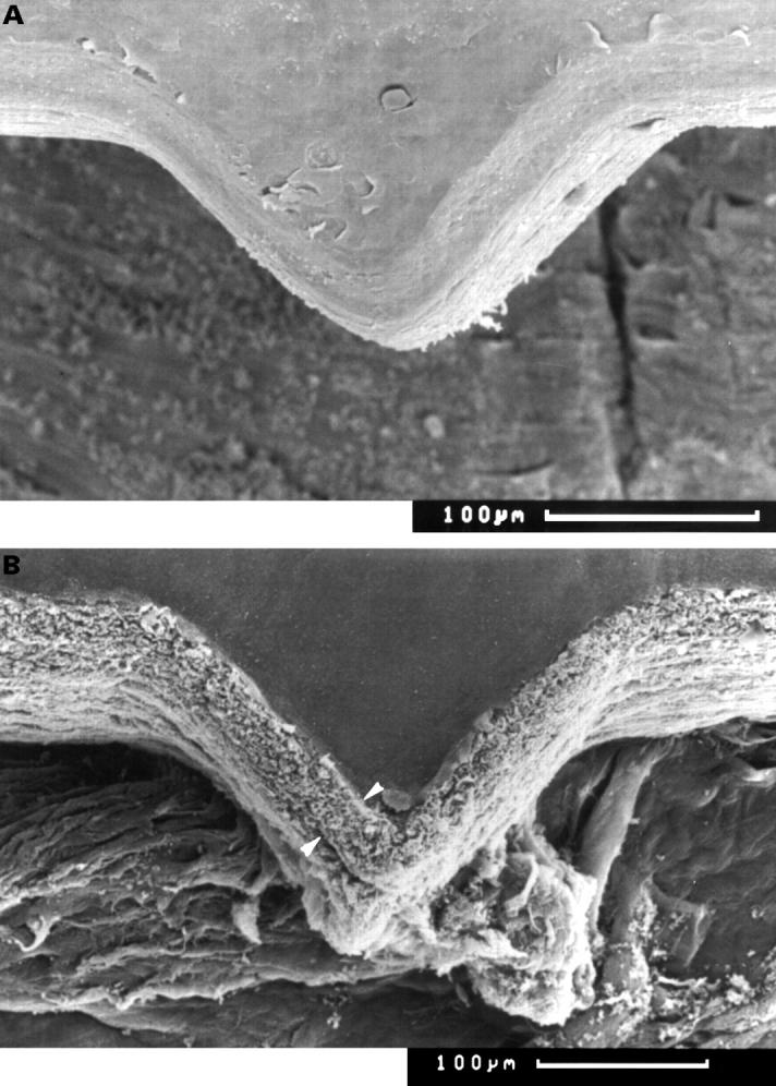
Figure 3 .

Scanning electron microscopy of "orientation teeth". (A) Excimer laser cut showing the triangular "orientation teeth" (×160). (B) Er:YAG cut of one orientation tooth at the margin of a donor cornea. An elevated surface of up to 35 µm can be observed (between arrowheads) (×150).
