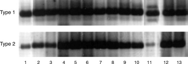
Figure 7 .
Presence of type 1 5α-reductase mRNA (Type 1) and type 2 5α-reductase mRNA (Type 2) in human ocular tissues and cells. Samples were processed for RT-PCR, agarose gel electrophoresis and ethidium bromide staining, as explained in Materials and methods. Photographs of agarose gels were obtained with a Polaroid camera (Polaroid Corporation, Cambridge, MA, USA) and images on the internegatives were then captured with a CCD-72S video camera (Hamamatsu Photonics, Japan), imported into Adobe Photoshop 4.01 and printed with a Kodak XLS 8600 printer. Samples 1-10 and 11-13 were run on parallel gels and bands have been aligned according to the molecular size of the bands. The numbers refer to the following samples: (1) male meibomian gland; (2) female meibomian gland; (3) female lacrimal gland; (4) male lacrimal gland; (5) prostate (surgical specimen); (6) Hs68 cells; (7) female conjunctiva (n = 3 combined tissues); (8) male conjunctiva (n = 3 combined tissues); (9) female cornea (n = 3 combined tissues); (10) male cornea (n = 3 combined tissues); (11) retinal pigment epithelial cells; (12) LNCaP cells; (13) prostate (RNA from Clontech).

