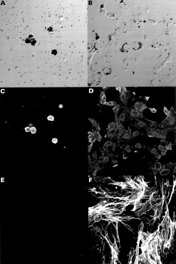
Figure 1 .
Photomicroscopy of retinal pigment epithelial (RPE) cells under bright field illumination (A, B), and immunofluorescent staining for cytokeratin (C, D) and α smooth muscle actin (α-SMA) (E, F) on day 0 (A, C, E) and day 14 of incubation with 100 ng/ml platelet derived growth factor (PDGF) (B, D, F). Freshly isolated RPE cells were hexagonal-shaped and mostly pigmented (A). RPE cells partly became flattened and lost their pigment granules (B). They exhibited a decline in immunoreactivity for cytokeratin (D) and an increase in α-SMA immunoreactivity (F). Original magnification, ×150.

